The human knee is one of the most complex joints in the body, playing a crucial role in movement, stability, and balance. Whether you’re walking, running, or standing, your knees bear the weight and pressure of your entire body. When pain, injury, or swelling occurs, one of the most common diagnostic tools used by doctors is a knee X-ray. But what does a normal knee X-ray actually look like? Let’s explore everything you need to know in this detailed guide.
What Is a Knee X-Ray?
A knee X-ray is a quick, non-invasive imaging test that uses low levels of radiation to create pictures of the bones and joint structures of the knee. It helps doctors evaluate the health of the bones and identify any abnormalities such as fractures, dislocations, arthritis, or infections.
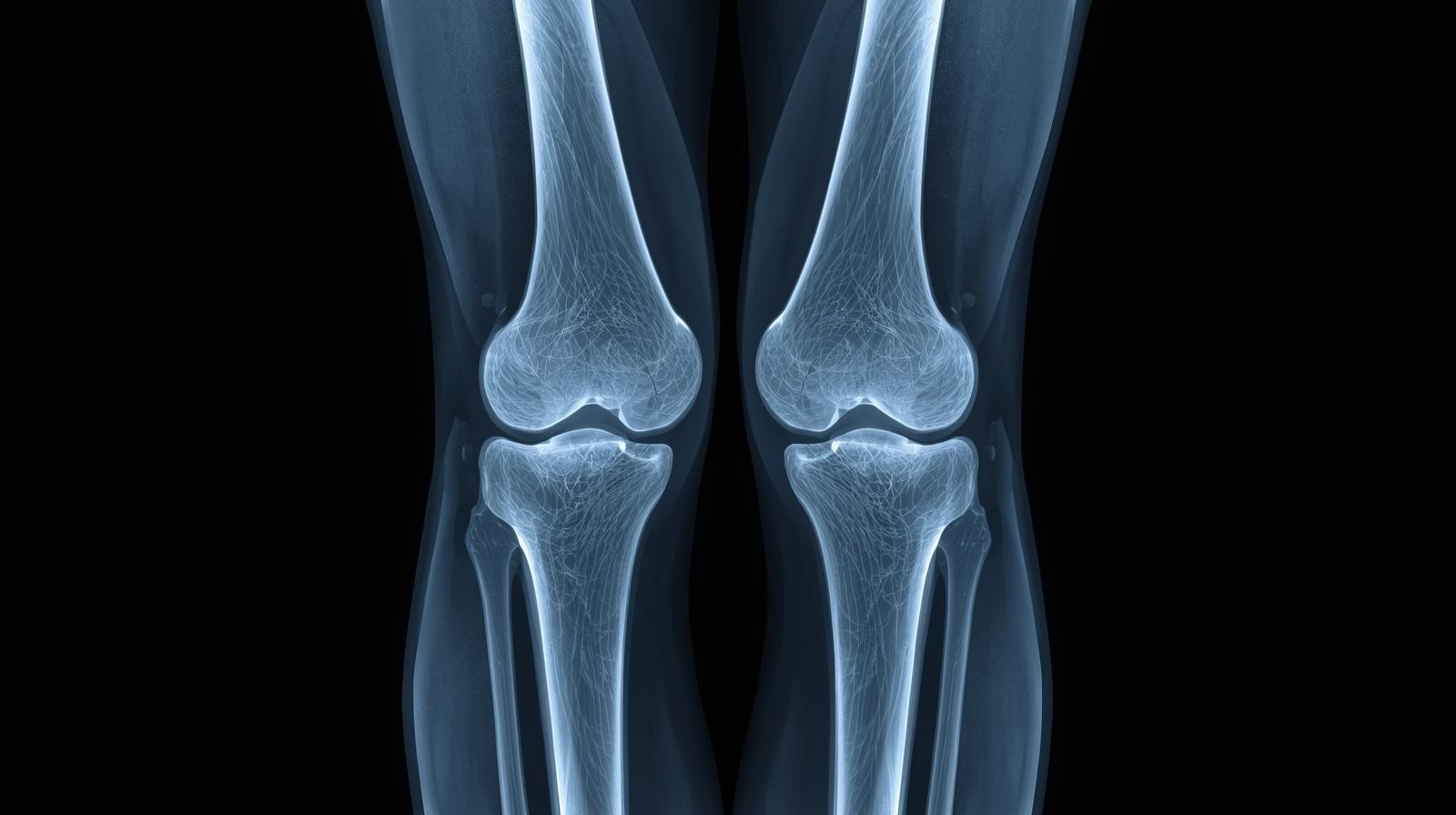
In a normal knee X-ray, the bones are properly aligned, the joint spaces are even, and there are no visible fractures or unusual bone growths. The X-ray can be taken from different angles to get a complete view of the joint. Common views include:
Anteroposterior (AP) View: This is a front-to-back image of the knee.
Lateral View: This shows the knee from the side.
Sunrise or Skyline View: Taken with the knee bent to show the kneecap and the groove it sits in.
These different views help doctors get a comprehensive understanding of the knee joint and surrounding bone structures.
Anatomy of the Knee Joint
To understand what a normal knee X-ray looks like, it’s helpful to know the basic anatomy of the knee. The knee joint consists of three major bones:
Femur (Thigh Bone): The upper leg bone that meets the lower leg bone at the knee.
Tibia (Shin Bone): The larger of the two lower leg bones that supports most of your body weight.
Patella (Kneecap): A small, triangular bone that protects the front of the knee joint.
These bones form two main articulations:
The tibiofemoral joint (between femur and tibia).
The patellofemoral joint (between patella and femur).
The joint is stabilized by ligaments including the anterior cruciate ligament (ACL), posterior cruciate ligament (PCL), and collateral ligaments while the cartilage acts as a cushion between bones.
What a Normal Knee X-Ray Shows
A normal knee X-ray displays clear, well-defined bone structures without any signs of damage, deformity, or misalignment. Here’s what doctors look for in a normal image:
Proper Joint Alignment: The femur, tibia, and patella are all in their correct positions.
Normal Joint Space: The space between the bones should be even, indicating healthy cartilage.
Smooth Bone Edges: The edges of the bones should be smooth without spurs or rough patches.
No Fractures or Cracks: The bone surfaces should appear continuous without any breaks.
Normal Bone Density: The bones should have consistent shading, showing healthy density and mineralization.
If the X-ray shows any narrowing of joint spaces, irregular bone surfaces, or abnormal shadows, it could indicate early signs of arthritis, cartilage wear, or other conditions.
How a Knee X-Ray Is Performed
Getting a knee X-ray is simple, fast, and painless. The procedure usually takes less than 10 minutes.
Preparation: The patient may be asked to remove any jewelry or clothing that could interfere with the image. A lead apron may be provided to protect other parts of the body from radiation.
Positioning: The radiologic technologist positions the patient depending on which views are needed.
Image Capture: The X-ray machine directs radiation toward the knee while an image detector captures the reflection.
Completion: The images are reviewed to ensure clarity before being sent to a radiologist for interpretation.
Indications for a Knee X-Ray
Doctors recommend a knee X-ray for many reasons, including:
Persistent pain or swelling in the knee
Difficulty moving the knee joint
A fall or accident causing trauma to the leg
Suspected fractures or dislocations
Chronic conditions like osteoarthritis or gout
Pre-surgical assessment or post-operative follow-up
In children, knee X-rays may be used to check bone growth and rule out developmental issues.
Understanding Normal vs. Abnormal Findings
When reviewing an X-ray, radiologists compare the findings with what’s considered a normal knee X-ray. Below are some examples:
Feature
Normal Knee X-Ray
Abnormal Knee X-Ray
Joint Alignment
Bones properly aligned
Bones displaced or misaligned
Joint Space
Even and well-defined
Narrowed or uneven (possible arthritis)
Bone Surface
Smooth edges
Rough edges or bone spurs
Fractures
None
Visible cracks or breaks
Patella
Centered in groove
Misaligned or dislocated
An abnormal knee X-ray might show osteophytes (bone spurs), decreased bone density, or fluid accumulation. Each of these findings gives clues about underlying knee conditions.
Limitations of Knee X-Rays
Although knee X-rays are excellent for detecting bone-related issues, they have limitations. They can’t visualize soft tissues like ligaments, tendons, or cartilage clearly. For these structures, doctors may order:
MRI (Magnetic Resonance Imaging): Best for soft tissues like the ACL or meniscus.
CT Scan: Offers detailed cross-sectional images of the knee.
Ultrasound: Useful for evaluating fluid buildup and tendon issues.
A normal X-ray doesn’t always mean the knee is problem-free pain may still originate from soft-tissue injuries not visible on the scan.
When to Seek Medical Advice
Even if your knee X-ray appears normal, it’s important to talk to your doctor if you have persistent knee pain, stiffness, or instability. These could indicate soft-tissue injuries that require further evaluation.
You should seek medical attention if you experience:
Severe knee swelling
Inability to bear weight
Locking or catching sensation
Redness or warmth around the knee
Your doctor might recommend additional tests, physical therapy, or medications based on your symptoms and X-ray results.
Preventing Knee Injuries and Maintaining Joint Health
Keeping your knees healthy can help you avoid injuries and the need for medical imaging. Some effective tips include:
Maintain a Healthy Weight: Extra body weight puts stress on your knees.
Exercise Regularly: Low-impact exercises like swimming, cycling, and walking strengthen muscles without straining joints.
Warm Up and Stretch: Before workouts, proper stretching helps prevent ligament injuries.
Wear Proper Footwear: Supportive shoes reduce impact on the knees.
Avoid Overuse: Give your knees rest if you experience discomfort during activities.
Following these steps can reduce your risk of knee problems and help maintain normal joint function over time.
Advances in Digital Knee X-Rays
Modern medical imaging has evolved significantly. Digital knee X-rays offer sharper, faster, and safer results compared to traditional film-based X-rays. They use less radiation, and the results can be stored electronically for easy sharing between healthcare providers.
Advanced image processing techniques also allow radiologists to zoom in, adjust contrast, and detect even minor bone changes. This technology ensures faster diagnosis and more accurate treatment planning.
Final Thoughts
A normal knee X-ray provides a clear picture of healthy bone alignment, even joint spacing, and smooth surfaces indicators of a stable and functional joint. While X-rays are primarily used to rule out fractures or bone diseases, they remain one of the most accessible and valuable tools in orthopedic care.
If your doctor says your knee X-ray is normal, that’s good news it means your bones are healthy and well-aligned. However, it’s still important to listen to your body and seek follow-up care if pain persists.
Maintaining strong muscles, flexible ligaments, and healthy bones will keep your knees functioning well for years. A normal knee X-ray is not just a clean report it’s a sign that your body’s most powerful joints are doing their job perfectly.