When it comes to diagnosing conditions involving the throat, neck, or airway, one of the most useful and commonly performed imaging tests is the soft tissue neck X-ray. This simple yet powerful test helps doctors visualize the soft tissues, bones, and airways in the neck region providing valuable insight into infections, obstructions, injuries, or foreign bodies.
The soft tissue neck X-ray is especially important because the neck houses several vital structures, including the trachea (windpipe), esophagus, thyroid gland, lymph nodes, and major blood vessels. Any swelling, infection, or blockage in these areas can cause serious symptoms like difficulty breathing or swallowing and that’s where X-rays play a crucial role in quick diagnosis.
Let’s take a detailed look at what a soft tissue neck X-ray is, when it’s needed, how it’s done, what doctors look for, and what patients should expect during and after the procedure.
What Is a Soft Tissue Neck X-Ray?
A soft tissue neck X-ray is a special type of radiograph that focuses on the soft structures of the neck rather than the bones alone. Standard X-rays primarily highlight dense materials like bones, which appear white on the image. However, with specific settings and exposure techniques, a soft tissue neck X-ray can also capture the outlines of the airway, trachea, tonsils, adenoids, and other soft tissue structures.
This imaging test allows doctors to detect abnormalities such as swelling, infections, or masses that may not be visible during a routine physical examination.
In medical settings, this X-ray is often ordered urgently when a patient presents with sudden breathing difficulty, severe sore throat, or signs of airway obstruction. It’s a quick and non-invasive way to visualize what’s happening inside the neck region.
Why Is a Soft Tissue Neck X-Ray Done?
There are several important reasons why a doctor might recommend a soft tissue neck X-ray. It’s used both in emergency situations and for general diagnostic purposes.
1. To Check for Airway Obstruction
If a patient is having trouble breathing, an X-ray can show whether the airway is narrowed or blocked. This might be due to swelling, inflammation, or a foreign object lodged in the throat.
2. To Detect Infections
Infections like epiglottitis, tonsillitis, or retropharyngeal abscess can cause the soft tissues of the neck to swell dangerously. A soft tissue neck X-ray helps identify these conditions early so that treatment can begin quickly.
3. To Locate Foreign Bodies
Children sometimes swallow small objects such as coins, buttons, or toys. Even adults can accidentally inhale or swallow food pieces that get stuck. The X-ray helps locate these foreign bodies and guide their removal.
4. To Evaluate Trauma
After an accident or injury to the neck, doctors use X-rays to look for fractures, swelling, or air leaks (subcutaneous emphysema) that could compromise breathing.
5. To Assess Tumors or Masses
If there’s suspicion of a growth, enlarged lymph node, or tumor in the neck, an X-ray can show abnormal shadows or enlargements in the soft tissue areas.
6. To Check the Position of Medical Tubes
In hospitals, patients who are intubated or have tracheostomy tubes often undergo soft tissue neck X-rays to confirm correct placement and ensure the airway remains clear.
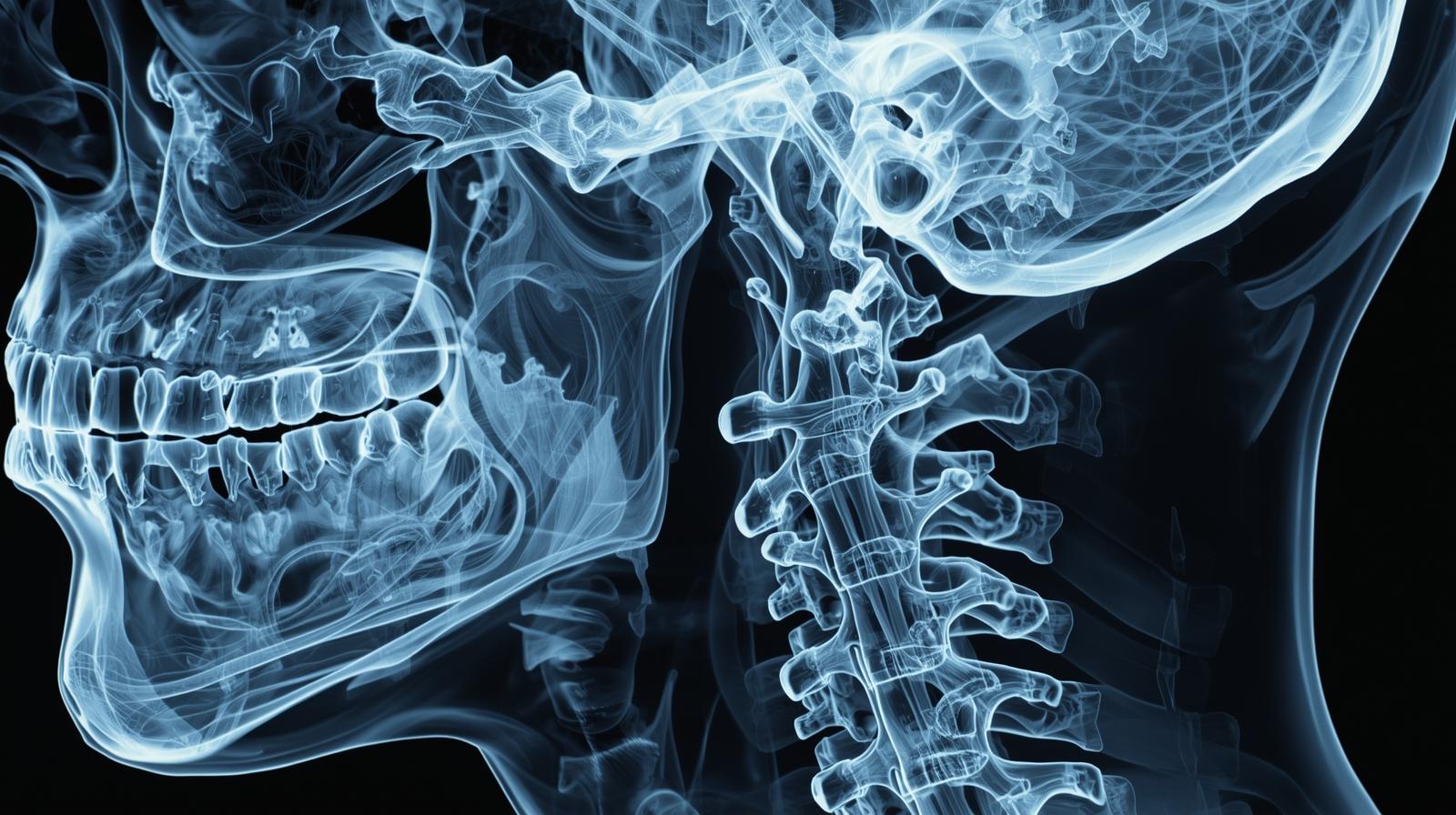
Anatomy Seen on a Soft Tissue Neck X-Ray
The neck is a complex area, and this X-ray provides a clear view of several key structures, including:
- Trachea (windpipe) – the main airway passage
- Epiglottis – a flap that closes over the trachea during swallowing
- Pharynx and larynx – throat and voice box regions
- Adenoids and tonsils – lymphatic tissues that can swell during infection
- Soft tissues – surrounding muscles and fat layers
- Cervical spine – upper vertebrae of the neck, which can affect nearby tissues
Radiologists and doctors analyze the X-ray carefully to identify any deviation, swelling, narrowing, or unusual appearance in these structures.
How the Soft Tissue Neck X-Ray Procedure Is Done
The procedure for a soft tissue neck X-ray is straightforward, quick, and painless. It can be done in a hospital’s radiology department or an urgent care facility.
Step 1: Preparation
- The patient may be asked to remove jewelry, eyeglasses, dentures, or any metallic objects that could interfere with the X-ray image.
- No special fasting or preparation is needed unless otherwise instructed.
Step 2: Positioning the Patient
The technologist will position the patient in front of the X-ray machine. Depending on the purpose of the scan, one or more of the following views may be taken:
- Lateral view (side view) – the most common angle for soft tissue neck X-rays.
- Anteroposterior (front-to-back view) – helps visualize tracheal alignment.
- Oblique view – sometimes used for more detail.
The patient may be asked to stand or sit upright and hold still during the exposure.
Step 3: Image Exposure
The technologist will briefly activate the X-ray beam, which passes through the neck to create the image. The entire process takes only a few seconds.
Step 4: Completion
Once the images are captured, the patient can resume normal activities immediately. A radiologist will review the images and send the report to the doctor who ordered the test.
What Does the Doctor Look for in the X-Ray?
Radiologists analyze several key features in a soft tissue neck X-ray, depending on the clinical question. Here are some common findings and what they mean:
1. Epiglottitis
This is a potentially life-threatening condition caused by inflammation of the epiglottis, often due to bacterial infection.
- On X-ray: The “thumb sign” appears a swollen epiglottis that looks like a thumb-shaped shadow.
2. Croup (Laryngotracheobronchitis)
Common in children, croup causes swelling in the upper airway.
- On X-ray: The “steeple sign” or “hourglass sign” is visible a narrow trachea that looks pinched.
3. Foreign Body
If a child or adult swallows something, the X-ray can show its location.
- On X-ray: The foreign object appears as a bright (white) shadow against the darker airway.
4. Retropharyngeal Abscess
A deep neck infection that can obstruct breathing.
- On X-ray: Widened prevertebral soft tissue area and sometimes gas pockets are visible.
5. Airway Deviation
If the trachea appears shifted to one side, it might indicate a tumor, enlarged thyroid, or pressure from another structure.
6. Subcutaneous Emphysema
When air leaks into the soft tissue after trauma or surgery.
- On X-ray: Air bubbles appear as dark streaks under the skin.
7. Tumors or Masses
Abnormal soft tissue shadows or asymmetry can indicate growths that require further imaging such as CT or MRI.
Safety and Radiation Concerns
A soft tissue neck X-ray involves a very small dose of radiation, which is considered safe for adults and children when used appropriately. Modern X-ray equipment uses advanced filters and digital systems to minimize exposure.
Doctors always weigh the benefits against the risks before recommending any X-ray. Pregnant women should inform their doctor before the test so protective shielding can be used if absolutely necessary.
In emergency situations such as airway obstruction the benefits of quick diagnosis far outweigh the minimal radiation risk.
How to Prepare for the Test
For most people, no special preparation is required. However, a few tips can help ensure accurate results:
- Avoid metallic accessories: Remove necklaces, earrings, or clothing with zippers or buttons near the neck.
- Stay still: Even slight movement can blur the image.
- Inform your doctor: Tell them about any past neck surgeries, implants, or known allergies to contrast agents (if any imaging contrast is used later).
After the X-Ray: What Happens Next?
Once the images are taken, the radiologist examines them and writes a detailed report. This is sent to your referring doctor, usually within a few hours or the same day.
Based on the findings, your doctor may:
- Prescribe antibiotics (for infections)
- Order a CT or MRI for detailed imaging
- Recommend removal of a foreign body
- Refer you to an ENT specialist (Ear, Nose, and Throat doctor)
If the results are normal, your doctor may continue with other tests to identify the cause of symptoms.
Advantages of Soft Tissue Neck X-Rays
- Quick and Non-Invasive: No needles or injections are required.
- Cost-Effective: Less expensive than CT or MRI scans.
- Widely Available: Can be done in most hospitals and clinics.
- Useful for Emergencies: Provides instant results in urgent cases.
- Guides Further Imaging: Helps determine if more advanced scans are needed.
Limitations of Soft Tissue Neck X-Rays
While very useful, X-rays do have some limitations.
- They provide less soft tissue detail compared to CT or MRI.
- Very small lesions or early infections might not be visible.
- Certain foreign bodies (like plastic) may not appear clearly.
In such cases, doctors may order additional imaging tests for confirmation.
Common Misconceptions About Soft Tissue Neck X-Rays
“It’s only for broken bones.”
No this specific X-ray focuses on soft tissues, airways, and possible infections, not just fractures.
“It’s dangerous because of radiation.”
Modern digital X-rays use extremely low doses of radiation often less than a single day’s exposure to natural background radiation.
“It always requires fasting.”
Fasting is not needed unless another test (like a contrast study) is planned on the same day.
“It’s painful.”
The procedure is completely painless and takes just a few minutes.
Case Example: Detecting a Foreign Body in a Child
A five-year-old boy came to the emergency room after swallowing a small coin. His parents noticed he was drooling and having difficulty swallowing. The doctor ordered a soft tissue neck X-ray immediately.
The image revealed the coin stuck at the upper esophagus level. Thanks to the quick X-ray, doctors removed it safely using an endoscope, avoiding surgery or complications.
This example highlights how valuable this test is in emergencies especially in children.
The Role of Radiologists and ENT Specialists
A radiologist is responsible for interpreting the X-ray images, identifying abnormalities, and providing a detailed report. The ENT specialist (ear, nose, and throat doctor) then uses this report to plan treatment or surgery.
Together, these professionals ensure that patients get accurate diagnoses and the right medical care promptly.
Advances in Digital X-Ray Technology
With digital radiography, soft tissue neck X-rays have become faster, safer, and clearer.
- Instant image viewing: Doctors can see results within seconds.
- Enhanced image quality: Allows zooming, contrast adjustment, and precise measurement.
- Reduced radiation: Digital sensors need less exposure.
- Easy sharing: Images can be sent electronically for second opinions.
These technological improvements have made soft tissue neck imaging even more reliable and efficient in both emergency and routine cases.
Key Takeaways
- A soft tissue neck X-ray is a quick, painless imaging test that examines the airway, throat, and surrounding tissues.
- It’s used to detect infections, swelling, foreign bodies, or airway obstruction.
- The procedure is safe, low in radiation, and ideal for emergency diagnosis.
- Radiologists interpret the images, and ENT specialists provide treatment based on the findings.
- Though it has some limitations, it remains a vital first-line diagnostic tool in neck-related medical conditions.
Final Thoughts
The soft tissue neck X-ray might seem like a simple test, but it often provides life-saving information. From identifying airway blockages to detecting serious infections, this diagnostic tool helps doctors act quickly and effectively.
For patients, the procedure is safe, fast, and painless often completed within minutes. For doctors, it’s an invaluable window into the complex anatomy of the neck.
As medical imaging technology continues to advance, the accuracy and clarity of these X-rays will only improve, helping healthcare professionals diagnose and treat conditions with even greater precision.
Whether used in emergency rooms, ENT clinics, or pediatric care, the soft tissue neck X-ray remains one of the most practical and reliable diagnostic tests in modern medicine simple yet powerful, saving lives one image at a time.